Ofer Reiter 1, Ilit Mimouni 2, Stephen Dusza 3, Allan C Halpern 3, Yael Anne Leshem 4, Ashfaq A Marghoob 3 Affiliations expand
- PMID: 31706938
- PMCID: PMC9366765
Free PMC article
Abstract
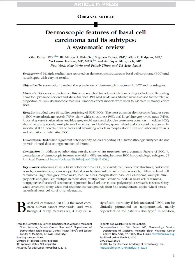
Background: Multiple studies have reported on dermoscopic structures in basal cell carcinoma (BCC) and its subtypes, with varying results. Objective: To systematically review the prevalence of dermoscopic structures in BCC and its subtypes. Methods: Databases and reference lists were searched for relevant trials according to Preferred Reporting Items for Systematic Reviews and Meta-Analyses (PRISMA) guidelines. Studies were assessed for the relative proportion of BCC dermoscopic features. Random-effects models were used to estimate summary effect sizes. Results: Included were 31 studies consisting of 5950 BCCs. The most common dermoscopic features seen in BCC were arborizing vessels (59%), shiny white structures (49%), and large blue-grey ovoid nests (34%). Arborizing vessels, ulceration, and blue-grey ovoid nests and globules were most common in nodular BCC; short-fine telangiectasia, multiple small erosions, and leaf-like, spoke wheel and concentric structures in superficial BCC; porcelain white areas and arborizing vessels in morpheaform BCC; and arborizing vessels and ulceration in infiltrative BCC. Limitations: Studies had significant heterogeneity. Studies reporting BCC histopathologic subtypes did not provide clinical data on pigmentation of lesions. Conclusion: In addition to arborizing vessels, shiny white structures are a common feature of BCC. A constellation of dermoscopic features may aid in differentiating between BCC histopathologic subtypes. Keywords: BCC; arborizing vessels; basal cell carcinoma; blue-white veil; concentric structures; corkscrew vessels; dermatoscopy; dermoscopy; dotted vessels; glomerular vessels; hairpin vessels; infiltrative basal cell carcinoma; large blue-grey ovoid nests; leaf-like areas; morpheaform basal cell carcinoma; multiple blue-grey dots and globules; multiple in-focus dots; multiple small erosions; nodular basal cell carcinoma; nonpigmented basal cell carcinoma; pigmented basal cell carcinoma; polymorphous vessels; rosettes; shiny white structures; shiny white-red structureless background; short-fine telangiectasia; spoke wheel areas; superficial basal cell carcinoma; ulceration.

. J Am Acad Dermatol. 2019 Nov 7

